
## Beyond the Spiral: How Science Became Art in a Binghamton University Conch Shell
Imagine a conch shell, its spiraling form a testament to nature’s artistry. Now picture it dissected, its intricate internal structure revealed in stunning detail. This isn’t just a scientific study – it’s a work of art.
More Than Just Images
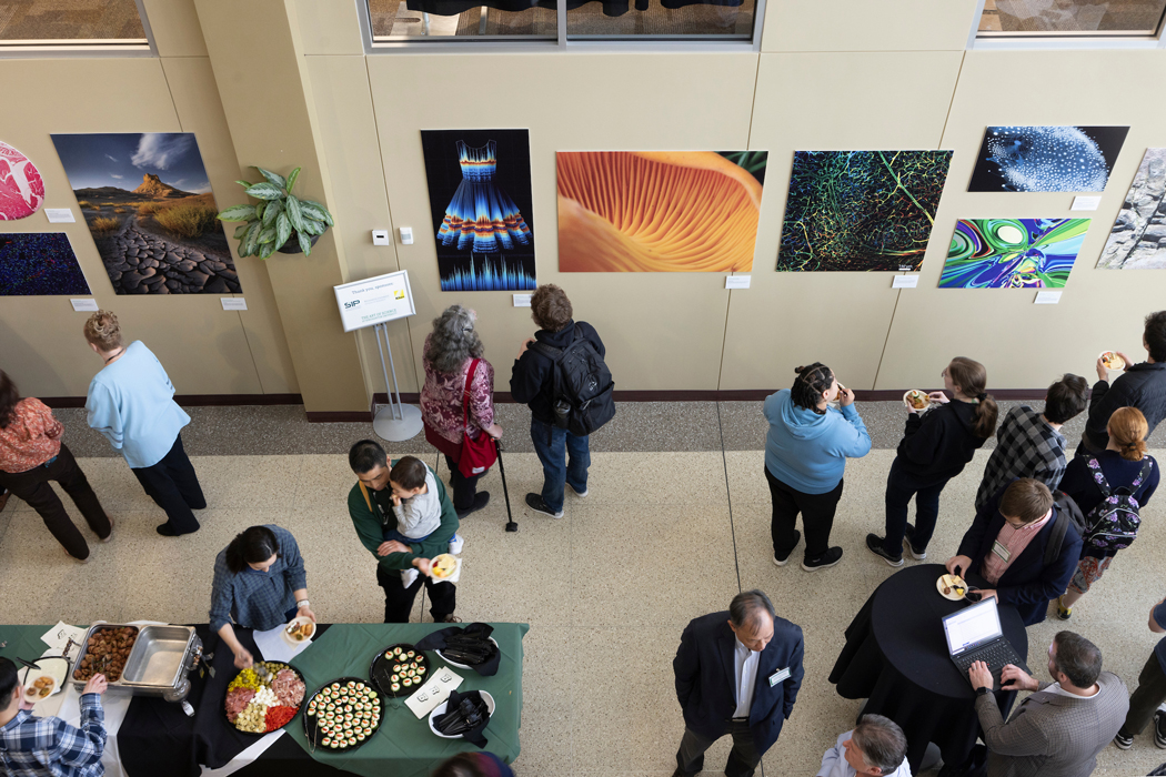
The Art of Science competition at Binghamton University is more than just a showcase of visually stunning images. It’s a platform that celebrates the inherent artistry and creativity found in scientific research. By encouraging researchers to present their work in innovative and engaging ways, the competition transcends the traditional boundaries of scientific communication.
The competition, organized by Martha Terry, assistant director of the Office of Research Advancement, includes two main categories: “The World Around Us,” featuring subjects visible to the naked eye, and “Visualizing the Unseen,” where instruments and optics are necessary to capture the subject matter. This dual approach highlights the diverse range of scientific inquiry happening at Binghamton University, from meticulous observations of nature to intricate explorations of the microscopic world.
The impact of Art of Science extends beyond the walls of the University. By showcasing the beauty and wonder hidden within scientific research, the competition inspires a wider audience to appreciate the interconnectedness of art and science. It sparks curiosity and encourages dialogue, fostering a deeper understanding of the complexities of the world around us.

Visualizing the Invisible
The “Visualizing the Unseen” category pushes the boundaries of conventional scientific illustration. Researchers utilize advanced imaging techniques and software to reveal hidden structures, processes, and phenomena that are invisible to the human eye. This category not only showcases the technical prowess of the researchers but also their ability to translate complex scientific data into compelling visual narratives.
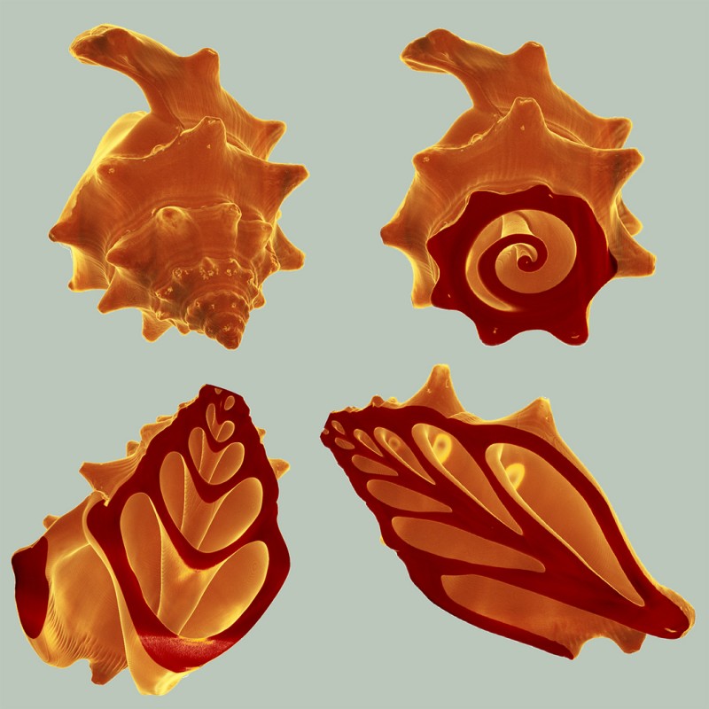
Anju Sharma, a senior scientist at the S3IP Center of Excellence, exemplifies this approach with her winning artwork, “Harmonicity in Nature.” Utilizing the Xradia Versa 620 X-ray microscope, Sharma captured intricate 3D images of a conch shell, revealing the Fibonacci spiral patterns that create a resonant chamber of harmonic frequencies. Her work demonstrates the power of X-ray microscopy to unveil the hidden beauty and complexity of natural structures.

Inspiring Innovation
Art of Science serves as a catalyst for innovation, inspiring both young scientists and artists to explore the intersection of their disciplines. By demonstrating the creative potential of scientific research, the competition encourages students to think outside the box and approach problems from unconventional angles.
The winning artworks often serve as springboards for further research and exploration. Researchers may discover new insights or connections within their data, leading to novel avenues of inquiry. The competition also fosters collaboration between researchers from different fields, as they share their expertise and perspectives.
Moreover, the public exposure generated by Art of Science can spark public interest in science and technology. By showcasing the beauty and wonder of scientific discovery, the competition can inspire a new generation of scientists, engineers, and innovators.

Unlocking Nature’s Secrets
The Power of X-ray Microscopy
X-ray microscopy, like the technique employed by Sharma, offers a unique window into the intricate world of biological structures, materials science, and beyond. By using X-rays to penetrate and image objects, researchers can visualize internal structures, processes, and compositions with unprecedented detail and clarity.
This non-destructive imaging technique allows scientists to study delicate samples without causing damage, preserving their integrity for further analysis. X-ray microscopy has revolutionized our understanding of biological systems, revealing the complex architectures of cells, tissues, and organisms.

Applications Across Disciplines
The applications of X-ray microscopy are vast and continue to expand. In medicine, it is used for diagnosing diseases, guiding surgical procedures, and monitoring treatment effectiveness. In materials science, it is employed to study the structure and properties of new materials, leading to advancements in fields such as electronics and energy production.
Here are just a few examples of how X-ray microscopy is being used to unlock nature’s secrets:
- Biology: Studying the intricate structures of viruses, bacteria, and other microorganisms, aiding in the development of new antibiotics and vaccines.
- Materials Science: Analyzing the microstructure of metals, ceramics, and polymers, improving their strength, durability, and performance.
- Geology: Examining the composition and structure of rocks and minerals, providing insights into the Earth’s history and geological processes.
- Archaeology: Non-destructively imaging ancient artifacts and structures, revealing hidden details and aiding in their preservation.

Pushing the Boundaries
The field of X-ray microscopy is constantly evolving, with ongoing advancements in technology pushing the boundaries of what is possible. Researchers are developing new techniques and instruments that offer even greater resolution, sensitivity, and versatility.
For example, advancements in X-ray sources and detectors are enabling the imaging of increasingly smaller and more complex structures. New computational methods are allowing scientists to process and analyze the vast amounts of data generated by X-ray microscopy, revealing hidden patterns and insights.
These ongoing developments promise to revolutionize our understanding of the world around us, unlocking new secrets in fields ranging from medicine and materials science to archaeology and astronomy.

A Window to the Unknown
X-ray microscopy is more than just a technological marvel; it is a powerful tool for expanding our understanding of the universe and our place within it. By peering into the hidden realms of matter and energy, we can gain a deeper appreciation for the complexity and interconnectedness of all things.
The ability to visualize the unseen opens up vast possibilities for scientific discovery, technological innovation, and artistic expression. As technology continues to advance, we can expect X-ray microscopy to play an increasingly important role in shaping our understanding of the world around us.

Conclusion
Unlocking the Hidden World Within: The Art of Science at Binghamton University
As we conclude our article on Binghamton University’s Art of Science winner, who shed light on the intricate details of a conch shell through an X-ray, it’s clear that this innovative approach to science communication has left an indelible mark. By using an X-ray to reveal the inner structures of the conch shell, the winner not only demonstrated their technical prowess but also highlighted the beauty and complexity of the natural world. This project serves as a testament to the power of interdisciplinary collaboration, where art and science converge to illuminate new perspectives and inspire a deeper appreciation for the world around us.

The significance of this project extends far beyond the confines of the university campus. It underscores the importance of making science accessible and engaging for a broader audience, and demonstrates the potential for innovative storytelling in science communication. As we move forward, it’s essential that we continue to push the boundaries of what is possible in science art, and that we provide opportunities for emerging talent to explore the intersection of art and science. By doing so, we can spark a new generation of thinkers, creatives, and innovators who will drive progress and discovery in the years to come.